-
New

-
New

-
New

-
New

-
New

-
New

-
New


Genoray
Genoray Vetera Ace C-Arm
Genoray Vetera Ace C-Arm
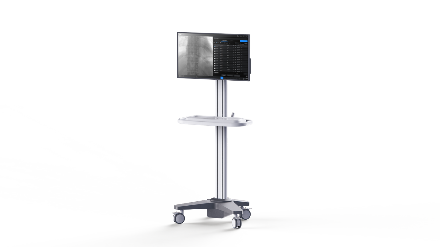
The Genoray VETERA ACE is an all-in-one mobile veterinary C-arm system with integrated FPD imaging, a compact body design, 860 mm free space, 3.2 kW HFG, and a 27" FHD touch monitor. It is designed specifically for veterinary fluoroscopy across a range of animal sizes.
Questions?


Collapsible content
Product Description
Genoray VETERA ACE Veterinary C-Arm System – All-in-One Mobile C-Arm with FPD
The Genoray VETERA ACE veterinary C-arm system is an all-in-one mobile fluoroscopy system designed specifically for veterinary use. Built with an integrated flat panel detector and compact body design, the VETERA ACE helps veterinary practices, specialty hospitals, and surgical centers perform real-time imaging procedures while optimizing limited procedure room space.
The system combines a 3.2 kW high-frequency generator, 860 mm free space, a 27" FHD touch monitor, and either a 20 x 20 cm CMOS or 21 x 21 cm TFT detector configuration. Its all-in-one design integrates key components without requiring an additional cart, supporting a cleaner and more efficient veterinary imaging workflow.
Key Features of the Genoray VETERA ACE
Veterinary-Specific C-Arm Design
The VETERA ACE is labeled for veterinary use only and is designed for animal imaging applications, making it distinct from Genoray’s human medical C-arm models.
All-in-One Mobile Platform
The integrated all-in-one body combines the main unit and key components without the need for a separate cart, helping maximize space in veterinary procedure rooms.
Integrated FPD Imaging
The system uses integrated flat panel detector technology to support modern digital fluoroscopy workflows for veterinary procedures.
860 mm Free Space
With 860 mm of free space, the VETERA ACE provides enhanced access for positioning animals and performing procedures with fewer spatial restrictions.
High-Speed Image Processing
Genoray’s low-latency image processing is designed to minimize image delay and support fast, accurate imaging during veterinary procedures.
Low-Dose Technology
The VETERA ACE includes low-dose technology intended to reduce radiation exposure while maintaining image quality for animals and users.
Advanced Imaging Workflow Tools
ZENIS IV Plus features include DNR, dose level adjustment, snapshot capture, real-time image comparison, image scroll, and style templates for customizable imaging preferences.
Optional DSA Capability
Digital Subtraction Angiography is listed as an available option, supporting contrast-enhanced fluoroscopic imaging when required.
Ideal Veterinary Applications
The Genoray VETERA ACE is well suited for:
- Veterinary fluoroscopy
- Orthopedic procedures
- Contrast-enhanced imaging
- Specialty animal hospitals
- Veterinary surgical centers
- Imaging across different species and animal sizes
Why Choose the Genoray VETERA ACE?
The VETERA ACE is a strong choice for veterinary teams that need a dedicated mobile C-arm with integrated FPD imaging, compact all-in-one construction, low-dose technology, and workflow tools tailored for efficient animal imaging. Its veterinary-specific design, large free space, and 27" touch monitor help support procedural accuracy in space-conscious clinical environments.
Key Benefits
- Designed specifically for veterinary use
- All-in-one mobile C-arm body
- Integrated FPD imaging
- 3.2 kW high-frequency generator
- 860 mm free space
- 27" FHD touch monitor
- Low-dose technology
- DNR and dose level adjustment
- Optional DSA capability
Specifications
GENORAY VETERA ACE
Veterinary Fluoroscope X-Ray System
Technical Specifications
SYSTEM OVERVIEW
• Mobile fluoroscopic X-ray system for veterinary use only
• Designed for fluoroscopic and digital spot imaging
• Intended for diagnostic, interventional, and surgical veterinary procedures
• Integrated C-arm gantry with detector and X-ray generator
• ZENIS IV Plus software platform
POWER REQUIREMENTS
• Rated Power: 200–230 V AC ±10%
• Power Consumption: 4.6 kVA
• Frequency: 50/60 Hz
• Maximum Supply Mains Impedance: ≤0.3 Ω
• Power Cable Length: 5 m
X-RAY GENERATOR
• Generator Type: High-frequency inverter
• Frequency: 38–44 kHz
• Phase: Single phase
• Nominal Peak Output Power: 3.2 kW
• kVp Range: 40–110 kV
Radiography Mode:
• 40–110 kV
• 1–100 mAs
Fluoroscopy – Low / Normal Mode:
• Continuous Fluoroscopy: 0.2–6.0 mA
• Pulsed Fluoroscopy: 1.0–20.0 mA
• Snapshot: 2.0–24.0 mA
Fluoroscopy – High Dose Mode:
• Continuous Fluoroscopy: 2.0–12.0 mA
• Pulsed Fluoroscopy: 2.0–24.0 mA
• Snapshot: 2.0–24.0 mA
MAXIMUM OPERATING DATA
Single Focus Type:
Radiography:
• 32 mA @ 100 kV
• 110 kV @ 16 mA
Fluoroscopy Low / Normal:
• Continuous: 6 mA @ 73 kV; 110 kV @ 4 mA
• Pulsed: 20 mA @ 49 kV; 110 kV @ 8.9 mA
• Snapshot: 24 mA @ 83 kV; 110 kV @ 18.1 mA
Fluoroscopy High Dose:
• Continuous: 12 mA @ 73 kV; 110 kV @ 8 mA
• Pulsed: 24 mA @ 83 kV; 110 kV @ 18.1 mA
• Snapshot: 24 mA @ 83 kV; 110 kV @ 18.1 mA
FLUOROSCOPY MODES
• Continuous fluoroscopy
• Pulsed fluoroscopy
• Pulse Rates: 30, 15, 8, 4, 2, and 1 pulse per second
• Snapshot mode
• Radiography mode
• Optional DSA
• Optional Road Map / RSA
DUTY CYCLE & EXPOSURE TIME
• Radiography Cooling Interval: 5 minutes between each X-ray exposure
• Fluoroscopy Duty Cycle: 1:60
• Maximum Fluoroscopy Exposure Time: 5 minutes
• Time Control Range: 0–5 minutes
FILTRATION & HVL
Total Filtration – Single Focus Type:
• 3.6 mm Al with 1.6 mm Al added filtration
• 4.8 mm Al with 2.8 mm Al added filtration
Total Filtration – Dual Focus Type:
• 3.6 mm Al with 2.0 mm Al added filtration
• 4.4 mm Al with 2.8 mm Al added filtration
HVL – Single Focus Type:
• 3.5 mm Al @ 80 kV with 1.6 mm Al added filtration
• 4.1 mm Al @ 80 kV with 2.8 mm Al added filtration
HVL – Dual Focus Type:
• 3.4 mm Al @ 80 kV with 2.0 mm Al added filtration
• 3.8 mm Al @ 80 kV with 2.8 mm Al added filtration
HOUSING HEAT UNIT
• Housing Heat Unit: 1,478,400 HU
X-RAY TUBE OPTIONS
D-068SBR Tube:
• Circuit: DC, center grounded
• Focal Spot: 0.6 mm
• Tube Type: Stationary
• Target Angle: 9°
• Maximum Tube Voltage: 110 kV
• Anode Heat Storage Capacity: 60 kJ / 84.5 kHU
OX/110-0514 Tube:
• Circuit: DC, center grounded
• Focal Spot: 0.5 / 1.4 mm
• Tube Type: Stationary
• Target Angle: 15°
• Cooling Time: 10 minutes
• Maximum Tube Voltage: 120 kV
• Anode Heat Capacity: 40 kJ / 56.4 kHU
X-RAY DETECTOR
20 x 20 Detector:
• Model: GFT-100
• Detector Type: CMOS
• Active Image Area: 201 x 200 mm
• Maximum Resolution: 3.3 lp/mm
• Frame Rate: 30 frames/sec
• Pixel Matrix: 1,344 x 1,320 pixels
• Pixel Sampling Resolution: 14 bits
• Pixel Pitch: 150 µm
• DQE: 50% @ 0.5 lp/mm
• MTF: 75% @ 0.5 lp/mm
21 x 21 Detector Option:
• Model: Mercu0909F
• Detector Type: a-Si TFT
• Active Image Area: 209.9 x 209.9 mm
• Maximum Resolution: 2.4 lp/mm
• Frame Rate: 30 frames/sec
• Pixel Matrix: 1,024 x 1,024 pixels
• Pixel Sampling Resolution: 16 bits
• Pixel Pitch: 205 µm
• DQE: 50% @ 0.5 lp/mm
• MTF: 80% @ 0.5 lp/mm
SYSTEM DIMENSIONS & WEIGHT
• Width: 800 mm
• Depth: 1,865 mm
• Height with Lift Movement: 1,808–2,308 mm
• Height with Monitor Arm Movement: 1,752–2,168 mm
• Weight: 310 kg ±5%
C-ARM GEOMETRY
• SID: 1,080 mm
• Free Space: 860 mm
• C-Arc Depth: 730 mm
• Vertical Range: 500 mm
• Horizontal Travel: 230 mm
• Orbital Rotation: 150°
• Lateral / Oblique Rotation: ±225° / 450° total
• Panning: ±15° / 30° total
• Lowest Lateral Height: 1,110 mm
MONITOR GEOMETRY
• Monitor Arm Swivel – First Arm: 200°
• Monitor Arm Swivel – Second Arm: 210°
• Monitor Arm Elevation Angle: 80°
• Monitor Arm Elevation Range: 416 mm
• Monitor Tilt: 20° total
• Monitor Swivel: ±95° / 190° total
• Lowest Vertical Height: 1,752–2,252 mm
OPERATION PANEL MOVEMENT
• OP Tilt:
– Upward: 20°
– Downward: 10°
• OP Swivel: 270°
COLLIMATOR
• Operation: Manual and remote motor drive
• Structure: Lead shutter
• 4-axis asymmetric lead shutter / square shutter
• 6-axis asymmetric lead shutter / octagonal iris shutter
• Virtual collimation
• Automatic collimation
• Adjustable shutter blades without X-ray exposure
Added Filter Options:
• 1.6 mm Al
• 2.0 mm Al
• 2.8 mm Al
LASER
• Type: Line beam laser diodes
• Laser Class: Class II
• Wavelength: 655 ±5 nm
• Power: DC 5 V ±5%
• Fan Ange: 58°
• Default Laser Duration: 10 seconds
• Maximum Adjustable Laser Duration: 30 seconds
IMAGE ACQUISITION FEATURES
• Fluoroscopy mode
• Radiography mode
• Snapshot mode
• LIH / Last Image Hold
• CINE image capture
• CINE save
• Image save
• Live image display
• Reference image display
• Thumbnail image review
• Manual kV adjustment
• Manual mA / mAs adjustment
• Automatic Brightness Control (ABC)
• FOV / zoom control
• Image calibration
• Import of previous or external images
IMAGE PROCESSING
• Dynamic Noise Reduction (DNR)
• Low / Normal / High DNR levels
• Motion Optimization
• Metal Object Optimization
• Brightness adjustment
• Contrast adjustment
• Sharpness adjustment
• Negative image
• Mirror image
• Upside-down image
• Image rotation
• Zoom in / zoom out
• Screen fit image
DOSE MANAGEMENT
• Low Dose mode
• Normal Dose mode
• High Quality / HLC mode
• Dose level adjustment
• DAP display
• CAK / cumulative air kerma display
• AKR / air kerma rate display
• Low Dose mode reduces dose rate by 50% or more compared to Normal Dose
• Normal Mode Dose Limit: <88 mGy/min
• High Dose / HLC Dose Limit: <176 mGy/min
• Radiation Dose Structured Report / RDSR support
ANIMAL / PATIENT SIZE PRESETS
• Patient size-based exposure settings
• Automatic adjustment of X-ray exposure conditions and image processing based on selected patient size
• Preset exposure tables for Low Dose, Medium Dose, and High Dose modes
ACCESSORIES
• Foot switch
• Hand switch
• Optional grid
External Cable Lengths:
• Power Cable: 5 m
• Hand Switch Cable: 0.8 m
• Foot Switch Cable: 5 m
Foot Switch Functions:
• Left Pedal: X-ray exposure
• Right Pedal: Image save
GRID
• Optional anti-scatter grid
• Designed to minimize scattered radiation
• Helps improve image clarity
• Detachable design
• Recommended to remove grid when using Low Dose mode
DISPLAY & SOFTWARE
• ZENIS IV Plus software platform
• Patient information management
• Image management
• DICOM-related workflow support
• Live image screen
• Reference image screen
• Equipment status display
• Patient information display
• Menu bar and reference image settings
CONNECTIVITY & I/O
• DisplayPort cable port
• USB port
• CD-ROM
• Ethernet network port
• DICOM network configuration support
• USB storage backup support
• External monitor connection support
SECURITY FEATURES
• User login required
• Admin, Standard, and CSE account roles
• Password expiration: 180 days
• Account locking and deletion support
• Screen saver security
• Firewall support
• Antivirus software support
• Closed network recommended
• DICOM service network limitation
SAFETY FEATURES
• Emergency stop switch
• X-ray key switch
• Brake system
• Indicator lamp
• Audible exposure alarm
• Exposure timeout protection
• Overheat warning
• Cooling status indication
• X-ray exposure ready status
• Error and emergency status display
Indicator Lamp Status:
• Lamp Off: Standby
• Green: X-ray exposure ready
• Yellow: X-ray exposure active
• Blue: Standby for X-ray exposure / cooling
• Red: Error or emergency stop
CLASSIFICATION & PROTECTION
• Class I medical equipment
• Equipment Water Resistance: IPX0
• Foot Switch Water Resistance: IPX7
• X-ray Tube Assembly Water Resistance: IPX2
• Laser Protection: Class 2
OPERATING ENVIRONMENT
• Operating Temperature: +15°C to +35°C
• Operating Humidity: 30% to 75% RH, non-condensing
• Operating Atmospheric Pressure: 800 to 1060 hPa
STORAGE ENVIRONMENT
• Storage Temperature: -15°C to +45°C
• Storage Humidity: 10% to 90% RH, non-condensing
• Storage Atmospheric Pressure: 700 to 1060 hPa
CLINICAL / VETERINARY APPLICATIONS
• Veterinary fluoroscopy
• Veterinary diagnostic imaging
• Veterinary surgical guidance
• Interventional veterinary procedures
• Digital spot imaging
• Orthopedic veterinary procedures
• Contrast studies
• Angiography with optional DSA / Road Map
FAQs
The Genoray Vetera ACE is an all-in-one mobile veterinary C-arm with an integrated flat panel detector (FPD) designed specifically for animal imaging. It provides real-time fluoroscopy and high-resolution imaging for a wide range of veterinary procedures, all in a compact, space-saving system.
What types of veterinary procedures is the Vetera ACE used for?The Vetera ACE supports a wide range of applications, including orthopedic surgery, trauma cases, pain management procedures, and contrast-enhanced fluoroscopy. It is designed for comprehensive veterinary imaging across different species and animal sizes.
Why should a veterinary practice choose the Genoray Vetera ACE?Veterinary clinics choose the Vetera ACE for its compact design, integrated FPD technology, low-dose safety features, and versatile imaging capabilities across animal types. It’s an ideal solution for practices looking to improve imaging performance while maximizing space and efficiency.
Additional Information
- Veterinary
- 21 cm
- Compact