-
Refurbished

-
Refurbished

-
Refurbished

-
Refurbished


GE Healthcare
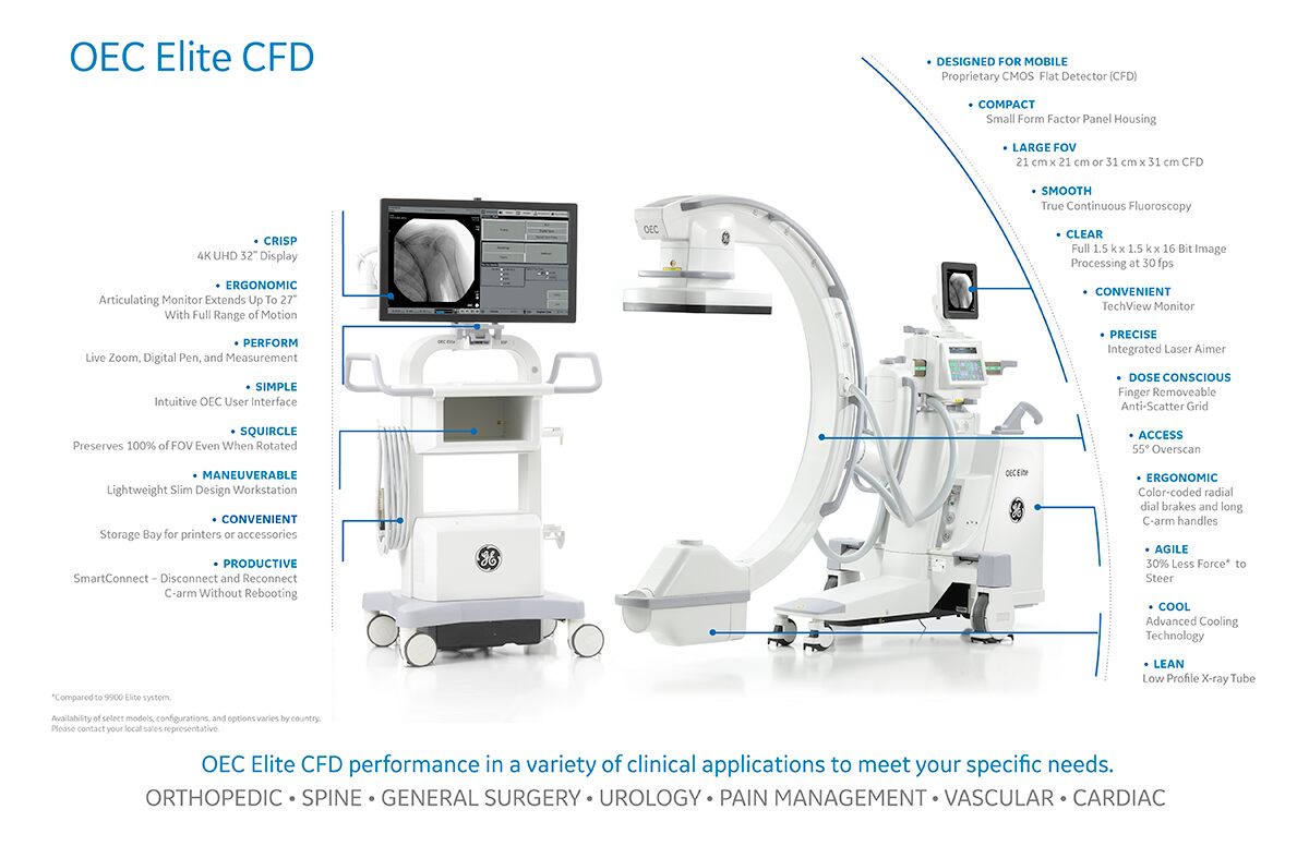
GE OEC Elite CFD C-Arm
GE OEC Elite CFD C-Arm
The GE OEC Elite CFD C-Arm System is a premium mobile surgical imaging system designed for orthopedic, spine, general surgery, urology, and pain management procedures. With proprietary CMOS flat detector technology, true continuous fluoroscopy, 1.5k x 1.5k imaging at 30 fps, and an intelligent workflow-focused design, it supports high-quality imaging with efficient positioning and dose-conscious operation.
Questions?


Collapsible content
Product Description
GE OEC Elite CFD C-Arm System – Premium Mobile Surgical Imaging with CMOS Flat Detector
The GE OEC Elite CFD C-Arm System is a premium mobile c-arm built for surgical teams that need advanced image quality, efficient workflow, and flexible positioning in the operating room. Designed with proprietary CMOS flat detector technology and an independent workstation, the system supports mobile surgical imaging across a wide range of procedures while helping clinicians see detailed anatomy with low-dose performance.
This system is designed to improve both imaging performance and everyday usability in the OR. It combines true continuous fluoroscopy, a compact detector head, an articulating touchscreen display, SmartConnect workflow capabilities, and enhanced dose control tools to support faster setup, easier maneuvering, and improved communication between the c-arm operator and surgical team.
Key Features of the GE OEC Elite CFD C-Arm System
CMOS Flat Detector Technology
The OEC Elite CFD features a proprietary CMOS flat detector with an ultra-efficient crystalline structure. The system is available with 21 cm x 21 cm and 31 cm x 31 cm flat detector options, giving facilities flexibility based on anatomy size and clinical application.
High-Resolution Imaging at 30 fps
The system provides full 1.5k x 1.5k x 16-bit image processing at 30 frames per second. It is designed to produce distortion-free images with strong anatomical detail and higher dynamic range for viewing structures with varying densities such as bone and soft tissue.
True Continuous Fluoroscopy
The OEC Elite CFD uses true continuous fluoroscopy rather than continuous pulse imaging. This helps capture more detailed anatomy without the visible lag, stutter, or ghosting that can distract from image interpretation during procedures.
Squircle Image Display Design
A distinctive squircle image display design preserves 100 percent of field of view even when the image is rotated. This helps clinicians maintain full image information on screen during procedures that require rotation and repositioning.
Super C-Arm Design with 55 Degree Overscan
The system features a deep Super C-arm design with 55 degrees of overscan, providing more free space and improved access around the patient. Its overall design also supports better visual line of sight and positioning in the surgical environment.
Improved Maneuverability and Positioning
The OEC Elite CFD is designed with updated steering and wheel design that requires 30 percent less force for positioning around the patient compared with other OEC models. An integrated laser aimer also helps operators position the detector precisely over the anatomy of interest.
Independent Workstation and SmartConnect Workflow
The lightweight slim workstation includes a flat work surface, accessory and storage bay, intuitive OEC user interface, physical keyboard, on-screen keyboard, and SmartConnect capability for disconnecting and reconnecting the c-arm without rebooting. This helps improve setup flexibility and productivity in active OR settings.
Articulating Touchscreen Monitor and TechView Display
The system includes a high-bright 27 inch LCD articulating display monitor with full range of motion and touchscreen controls for comfortable viewing. A TechView monitor is also included to improve communication with the surgical team during procedures.
Enhanced Dose Control Features
The OEC Elite CFD includes selectable default dose settings, High Level Fluoro or Digital Spot default pedal options, dose reports viewable on the workstation even when the c-arm is disconnected, RDSR access through DICOM or XML export, and a removable anti-scatter grid to help minimize dose for smaller patients.
Advanced Cooling and Low Profile Tube Design
The system includes advanced cooling technology and a low profile x-ray tube to support demanding surgical imaging workflows while maintaining a compact, mobile form factor.
Ideal Clinical Applications
The GE OEC Elite CFD C-Arm System is well suited for:
- Orthopedic surgery
- Spine procedures
- General surgery
- Urology
- Pain management procedures
Why Choose the GE OEC Elite CFD C-Arm System?
The GE OEC Elite CFD is a strong choice for facilities looking for a premium mobile c-arm with flat detector imaging, advanced workflow tools, and dose-conscious design. Its combination of CMOS detector technology, true continuous fluoroscopy, intelligent workstation features, and maneuverability improvements makes it well suited for surgical teams that need both imaging performance and operational efficiency.
Specifications
X-Ray System:
Generator:
- 60kHz high frequency
- 15kW power
- Up to 120kVp
- Up to 75mA for radiographic film exposure
- Continuous high level fluoro (HLF) up to 20mA
- Digital spot up to 75mA
- Pulsed HLF up to 40mA
- Full power from standard wall outlet
- Patented battery buffered design
X-Ray Tube:
- Rotating anode X-ray tube
- 0.3mm and 0.6mm focal spots
- Anode heat capacity: 300,000 HU
- Anode cooling rate: 85,000 HU/min.
- Housing heat capacity: 1,600,000 HU
- Housing cooling: -9″ II = 22,500 HU/min, 12″ II = 22,500 HU/min
Digital Image Orientation:
- Digitally adjusts image display for live and last image hold
- Automatic image update preserves image orientation settings applied during live and last image hold subsequent images
– Image rotation
– Image reversal (side-to-side)
– Image invert (top-to-bottom) - On-screen orientation indicator
- Fully digital with precise 1 degree rotation increments
PreView™ Collimator:
- On-screen collimator position indication
- PreView™ iris collimator
- PreView™ Tungsten rotatable double leaf collimator
- Adjusts collimators without X-ray exposure
Fluoro Mode:
- kVp range: 40 -120
- mA range: 0.2 – 10 normal mode 0.2 – 20 HLF (high level fluoro)
- Auto and manual fluoro modes
Pulsed Fluoro Mode:
- kVp range: 40 – 120
- mA range: 0.2 – 10 normal mode, 0.2-40 HLF
- Pulse rate: 8 pps
- Pulse width: 25 ms
- Auto and manual pulsed fluoroscopy modes
- Reduces x-ray dose to patient and operator
Digital Spot Mode:
- kVp range: 40 – 120
- mA range: Up to 75
- Automatic exposure termination and automatic image save
Video Imaging System:
21 cm CMOS Flat Panel Detector (CFD):
- Tri-mode 21 cm/15 cm/11 cm
- Minimum central resolution (at display):
– 21 cm: 2.5 lp/mm
– 15 cm: 3.0 lp/mm
– 11 cm: 3.3 lp/mm - Matrix: 1536 x 1496 total pixels
- DQE(0): 72%
- Pixel Pitch: 135.3 μm
- Removable grid with on-screen detection status
31 cm CMOS Flat Panel Detector (CFD):
- Tri-mode 31 cm/21 cm/15 cm
- Minimum central resolution (at display):
– 31 cm: 1.8 lp/mm
– 21 cm: 2.2 lp/mm
– 15 cm: 2.4 lp/mm - Matrix: 1548 x 1524 total pixels
- DQE(0): 72%
- Pixel Pitch: 198.0 μm
- Removable grid with on-screen detection status
Precision imaging with Dynamic Range Management (DRM) enhances features of interest while attenuating background noise:
- Preset Imaging Profiles
– 9800
– General
– Orthopedic
– Spine
– Cardiac
– Vascular
– Bolus Chase
AutoTrak™ Automatic Brightness Stabilization (ABS)
- Automatically seeks the subject anatomy anywhere within the imaging field
- Selects the optimum imaging technique by varying mA, kVp, and gain
- Automatically adjusts to anatomical size and location
- Provides uniform image quality throughout the entire image
Image I.Q.:
- Smart Window
– Dynamically senses the collimator position and automatically adjusts brightness and contrast to produce high image quality. - Smart Metal
– Allows user to adjust automatic brightness and contrast sensitivity levels to metal
– Provides optimum image quality even when metal is introduced to the field - Tungsten Collimator
– Denser collimator limits X-ray exposure area
– Reduces scatter radiation
– Improves image detail
Image Processing:
- 1k x 1k x 16 bit image intensifier
Video Monitor:
- 27 in (69 cm) LCD TFT color display
- Anti-glare
- Monitor mounted on an articulating arm
– 45 in (114 cm) horizontal travel
– 17 in (43.2 cm) vertical travel
– 27 in (68.8 cm) forward travel
– 5° up/5° down tilt
– Display viewable from all four sides of workstation
– Horizontal and vertical viewing angle 170° - 450 cd/m2 maximum brightness
- Touch screen system control
- 2560 x 1440 high resolution display
- Integrated PIP window to display color DVI-D input
FAQs
In the GE OEC Elite CFD, “CFD” refers to the system’s flat detector design, which GE highlights as a key reason the unit can provide excellent image detail at low dose. GE positions the Elite CFD as a premium flat detector C-arm for facilities that want strong visualization, workflow efficiency, and broad clinical versatility.
What are the main imaging benefits of the OEC Elite CFD?The OEC Elite CFD is designed to provide high image clarity, detailed visualization, and low-dose imaging support. GE highlights features such as Live Zoom, advanced noise reduction, and preset imaging profiles to help clinicians visualize anatomy and devices more clearly during procedures. The system also offers a 4K UHD display for improved viewing in the surgical environment.
How does the GE OEC Elite CFD help with field of view and positioning?One of the major advantages of the GE OEC Elite CFD C-arm is its ability to show up to 22% more field of view compared with monoblock C-arms, according to GE. GE also emphasizes its ergonomic design, low-profile X-ray tube housing, and the ability to reach difficult angles and low table heights more easily, which can help improve positioning around the patient during surgery.
Additional Information
- Orthopedics
- Pain Management
- Urology
- Vascular
- Veterinary
- 21 cm
- 31 cm
- Full Size