-
New

-
New

-
New

-
New

-
New

-
New

-
New

-
New


Fuji
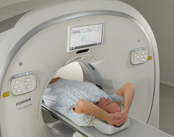
Fuji Scenaria View CT
Fuji Scenaria View CT
A premium multi-slice CT system engineered for high-speed acquisition, exceptional image clarity, and advanced low-dose performance, featuring intelligent workflow automation and powerful image reconstruction technologies. Designed to support a broad spectrum of clinical applications, the Scenaria View delivers reliable, high-throughput diagnostic imaging with enhanced patient comfort and operational efficiency for modern radiology departments.
Questions?


Collapsible content
Product Description
Fujifilm SCENARIA View CT – Advanced 128-Slice Performance with Intelligent Imaging
The Fujifilm SCENARIA View CT is a high-performance 128-slice computed tomography system engineered to deliver exceptional image quality, rapid acquisition speeds, and optimized dose efficiency. Designed for hospitals and advanced imaging centers, the SCENARIA View combines powerful hardware, intelligent automation, and patient-centered design to support a full range of diagnostic applications.
Built on Fujifilm’s advanced imaging platform, the SCENARIA View CT scanner provides superior spatial resolution and fast reconstruction capabilities — helping clinicians achieve confident diagnoses while improving workflow efficiency.
Key Features of the Fujifilm SCENARIA View CT
-
128-Slice Acquisition Capability
High-speed imaging enables detailed visualization for complex studies, including cardiac and vascular imaging. -
Advanced Iterative Reconstruction Technology
Reduces image noise while maintaining clarity, allowing for lower radiation dose without compromising diagnostic quality. -
Fast Gantry Rotation Speed
Improves temporal resolution and reduces motion artifacts, ideal for cardiac and emergency imaging. -
Wide Bore Gantry Design
Enhances patient comfort and accommodates larger or bariatric patients. -
High-Performance Detector & Generator System
Ensures consistent, high-resolution imaging across a broad range of clinical protocols. -
Optimized Dose Management Tools
Intelligent exposure control helps balance radiation reduction with image precision. -
Streamlined Workflow Automation
Simplified protocol selection and user-friendly interface reduce exam setup time and increase throughput. -
Seamless DICOM Connectivity
Integrates smoothly with PACS, RIS, and hospital IT infrastructure.
Clinical Applications
The Fujifilm SCENARIA View CT is ideal for:
- Cardiac CT & Coronary Imaging
- Neurological & Brain Studies
- Chest & Lung Imaging
- Abdominal & Pelvic Scans
- Trauma & Emergency Imaging
- Oncology Staging
- CT Angiography (CTA)
Its 128-slice capability makes it especially valuable for advanced cardiac and vascular applications requiring high temporal resolution.
Why Choose the Fujifilm SCENARIA View CT?
The SCENARIA View CT delivers:
- Advanced 128-slice performance
- Enhanced cardiac imaging capability
- Intelligent dose reduction technology
- Rapid throughput for busy imaging departments
- Compact yet powerful system design
Backed by Fujifilm’s global imaging expertise, the SCENARIA View provides a balance of speed, clarity, patient comfort, and operational efficiency.
Elevate Your Diagnostic Imaging with the Fujifilm SCENARIA View CT
If you're seeking a high-performance 128-slice CT scanner that offers advanced cardiac capability and optimized workflow, the Fujifilm SCENARIA View CT is a powerful and future-ready solution.
Specifications
FUJIFILM SCENARIA® VIEW CT SYSTEM
Technical Specifications
SYSTEM OVERVIEW
- Up to 128-slice capability
- 0.35 second minimum rotation time
- 80 cm wide bore gantry aperture
- 4 cm detector coverage (64 rows)
- Advanced dose reduction technologies
- Compact three-module system (gantry, table, console)
- Meets and surpasses Smart Dose Standard XR-29
SCAN PARAMETERS
- Rotation Times (sec): 0.35, 0.4, 0.5, 0.75, 1.0, 2.0
- Data Collection Speed: 2880 views per second
- Minimum Slice Thickness: 0.625 mm
- Field of View (FOV): 20 – 500 mm
- Maximum Scan Range: 79 in (2000 mm)
- Maximum Scan Rotations: 100
- Volume Scan Pitch: 0.578 – 1.578
SCAN ACQUISITION TYPES
- Scanogram (AP & Lateral) with real-time display
- Axial mode
- Helical (Volume) mode
- Dynamic Scan mode (Time Density Analysis)
- ECG Scan modes – Retrospective & Prospective (optional)
- Shuttle Scan mode (80 mm axial, 120 mm helical options)
GANTRY
- Aperture: 80 cm
- Gantry Tilt: ±30°
- 15" LCD Touchscreen Gantry Display
- ECG Data Input
- Laser localizer (prep & scan positions)
- Integrated table & gantry controls
- Patient intercom and auto-voice
- Breathing lights
DETECTOR
- Type: Solid-state ceramic
- Coverage: 4 cm with 64 rows
- Detector Elements: 64 x 888
- High-voltage design with up to 25% less electronic noise
X-RAY TUBE
- Anode Heat Capacity: 7.5 MHU
- Equivalent Heat Capacity: 45 MHU with Intelli IPV
- Max Cooling Rate: 1,386 kHU/min
- Tube Cooling: Oil/Air
- Focal Spots: 0.7 x 0.8 mm; 1.2 x 1.4 mm
- No external chiller required
X-RAY GENERATOR
- Type: High-frequency inverter control
- Output: 84 kW
- kVp Selection: 80 / 100 / 120 / 140 kV
- mA Selection: 10 – 700 mA (5 mA steps)
- Max Tube Current: 700 mA @ 100 & 120 kV
PATIENT TABLE
- Table-top Width: 18 in (475 mm)
- Table Length: 110 in (2805 mm)
- Weight Capacity: 660 lbs (299 kg)
- Minimum Table Height: 19 in (490 mm)
- Maximum Table Height: 38 in (970 mm)
- Horizontal Accuracy: ±0.25 mm
- Horizontal Travel Range: 83 in (2110 mm)
- Table Speed: 5 – 200 mm/sec
- Lateral Shift Range: ±10 cm (20 cm total range)
- Lateral Shift Speed: 10 mm/sec
IMAGE RECONSTRUCTION
- 128-slice per rotation capability
- Slice Thickness: 0.625 – 10 mm
- Reconstruction Speed: Up to 60 images/sec
- CT Number Range: -32,768 to +32,767
- Matrix: 512 x 512
- Up to 1024 x 1024 display matrix
DOSE MANAGEMENT TECHNOLOGIES
- Intelli IPV (Iterative Reconstruction)
– Up to 83% dose reduction vs FBP
– Up to 90% noise reduction
– Maintains natural image texture - Intelli EC Plus (3D mA modulation)
- CT Dose Check (XR-25 compliant)
- DICOM Dose Structured Report (XR-29 compliant)
- Simple Dose Report
- Multi-Bowtie Filters
- Reduced kV Imaging (80, 100, 120, 140 kV)
- Predict Scan (automated contrast timing)
- HiMAR Plus (Metal Artifact Reduction)
CARDIAC PACKAGE (OPTIONAL)
- Prospective ECG Gating
- Retrospective ECG Gating
- ECG Dose Modulation
- Cardio Conductor (protocol automation)
- Cardio Harmony (phase selection automation)
- Gate Editor
OPERATOR CONSOLE
- 24" LCD widescreen monitor
- Resolution: 1920 x 1200
- Dual Intel CPUs
- Windows 10 OS
- Fully automated, protocol-driven workflow
- Preview & Priority reconstruction
- Auto-archive and auto-film
- Exam-split functionality
- UPS included
3D POST-PROCESSING
- Multi-Planar Reconstruction (MPR)
- MIP / MinIP
- Surface Rendering
- Volume Rendering
- Auto MPR
STORAGE CAPACITY
- Image Storage: >1 TB (≥600,000 images)
- Raw Data Storage: 3 TB (>6,000 scan rotations)
- Archive: DVD-R / CD-R
NETWORK CAPABILITY
- IHE-SWF
- IHE PDI
- DICOM Modality Worklist (MWM)
- DICOM MPPS
- DICOM Query/Retrieve
- DICOM Dose SR
- Multi-destination image series send
POWER & ENVIRONMENTAL REQUIREMENTS
- Input Voltage: 480 VAC, 3 Phase
- Main Breaker: 200 AMP
- Peak Supply Capacity: 100 kVA
- Input Frequency: 50/60 Hz
- Power Regulation: ±5%
- Voltage Fluctuation: ±10%
- Operating Temperature: 20–28°C
- Operating Humidity: 35–80%
- Water Supply: Not required
PHYSICAL SPECIFICATIONS
Gantry
- Height: 2000 mm
- Depth: 943 mm
- Width: 2350 mm
- Weight: 2220 kg
Patient Table
- Height: 490 – 940 mm
- Depth: 2803 mm
- Width: 650 mm
- Weight: 500 kg
Operator Console
- Height: 606 mm
- Depth: 745 mm
- Width: 421 mm
- Weight: 74 kg
- Minimum Floor Plan: 285 sq. ft.
- Typical Floor Plan: 510 sq. ft.
FAQs
The Fujifilm Scenaria View CT scanner is designed as a multi-slice CT system engineered for high-resolution volumetric imaging. Depending on system configuration, it supports advanced slice acquisition suitable for comprehensive diagnostic imaging. The detector architecture is optimized for spatial resolution and consistent image uniformity across neuro, thoracic, abdominal, and musculoskeletal applications. Radiologists evaluating the Scenaria View typically consider it for its balance of acquisition speed, image clarity, and workflow efficiency.
How does the Fujifilm Scenaria View handle image reconstruction and post-processing?The Fujifilm Scenaria View CT system supports advanced image reconstruction techniques, including multiplanar reformatting (MPR), maximum intensity projection (MIP), and 3D volume rendering. Reconstruction speed is designed to support high-throughput environments while maintaining image fidelity. Its processing platform allows radiologists to review studies in axial, coronal, and sagittal planes with consistent clarity. Post-processing capabilities depend on the installed workstation and software configuration.
Does the Fujifilm Scenaria View CT scanner support cardiac imaging?Yes, depending on configuration. The Fujifilm Scenaria View CT system can support cardiac CT and coronary CTA applications when equipped with the appropriate slice configuration and cardiac software packages. Acquisition speed and temporal resolution are key considerations for facilities planning to perform cardiac studies, and system capabilities should be reviewed based on the specific installed model.
Additional Information
- Cardiac
- Pulmonary
- Oncology