Skip to content
Skip to product information
1 of 6

Fuji

Fuji OASIS Velocity MRI

Fuji OASIS Velocity MRI

The Fuji Oasis Velocity is a 1.2T high-field open MRI system designed to combine patient comfort with advanced diagnostic imaging performance. Featuring Synergy DLR deep learning reconstruction, IP-RAPID accelerated imaging, and a true open architecture, Oasis Velocity helps imaging centers improve scan efficiency, image quality, and patient accessibility.

Request A Quote View full details

Collapsible content

Product Description

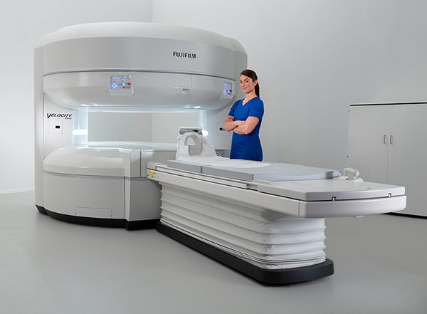
Fuji Oasis Velocity 1.2T Open MRI System – High-Field Open MRI with AI Reconstruction

The Fuji Oasis Velocity MRI System is a 1.2T high-field open MRI designed for imaging centers that want the comfort and accessibility of open MRI with the performance capabilities needed for a broad range of diagnostic exams. Its true open architecture helps reduce patient anxiety while improving access for bariatric, pediatric, elderly, claustrophobic, and mobility-limited patients.

Built with Synergy DLR deep learning reconstruction and IP-RAPID accelerated imaging, the Oasis Velocity is designed to reduce scan times while maintaining excellent image quality. The system supports high-resolution imaging across neuro, body, orthopedic, breast, spine, and musculoskeletal applications, making it a versatile MRI solution for modern outpatient imaging centers and hospitals.

Key Features of the Fuji Oasis Velocity

1.2T High-Field Open MRI Design
The Oasis Velocity combines a 1.2T high-field magnet with an open-sided design, giving facilities a patient-friendly MRI platform without sacrificing broad clinical capability.

Synergy DLR Deep Learning Reconstruction
Synergy DLR uses deep learning reconstruction to reduce image noise and support shorter exams with excellent image quality.

IP-RAPID Accelerated Imaging
IP-RAPID combines parallel imaging, sparse sampling, and iterative processing to help shorten scan times, boost resolution, and improve workflow efficiency.

Patient-Friendly Open Architecture
The open design helps patients feel less confined during exams and allows caregivers or technologists to remain close to pediatric, anxious, or mobility-challenged patients.

Wide, Accessible Patient Table
The table supports up to 660 lb, lowers to 20 inches for easier access, and includes a 32-inch wide design for comfortable positioning.

Advanced RF Coil Technology
The system includes sensitive multichannel RF coil technology, integrated head and spine coils, and blanket coil options designed to support patient comfort, coverage, and workflow.

3-Axis Motorized Lateral Table Movement
Motorized lateral table movement helps simplify iso-center positioning for extremity, spine, body, and musculoskeletal imaging.

SoftSound Gradient Technology
SoftSound gradient technology helps reduce acoustic noise, creating a more comfortable experience for patients during MRI exams.

Ideal Clinical Applications
The Fuji Oasis Velocity MRI system is well suited for imaging centers and hospitals performing a wide range of MRI exams, including:

  • Brain and neuro imaging
  • Spine imaging
  • Orthopedic and MSK imaging
  • Breast imaging
  • Body imaging
  • Pediatric MRI
  • Bariatric patient imaging
  • Claustrophobic or anxious patient exams


Why Choose the Fuji Oasis Velocity?

The Fuji Oasis Velocity open MRI system gives facilities a strong combination of patient comfort, high-field imaging performance, and workflow efficiency. With its true open design, AI-powered reconstruction, accelerated imaging, and accessible table design, Oasis Velocity is an excellent option for imaging providers that want to serve a broader patient population while improving throughput and image quality.

Key Benefits

  • 1.2T high-field open MRI platform
  • True open architecture for patient comfort
  • Synergy DLR AI reconstruction supports shorter exams
  • IP-RAPID helps improve speed and resolution
  • Wide table with 660 lb capacity
  • Designed for neuro, MSK, spine, body, and breast imaging

Specifications

FUJIFILM OASIS VELOCITY
1.2T Open MRI System

Technical Specifications

SYSTEM OVERVIEW

• 1.2T open superconducting MRI system
• Open vertical field magnet design
• Designed for high-field imaging with improved patient comfort
• Unobstructed lateral patient viewing angle
• Built for broad patient demographics, including larger and claustrophobic patients
• Includes Synergy DLR AI reconstruction and IP-RAPID enhanced image reconstruction

MAGNET SYSTEM

• Magnet Type: Superconducting open magnet
• Field Strength: 1.2T
• Field Orientation: Vertical field
• Homogeneity: 0.3 ppm @ 35 cm DSV
• Active magnetic shielding
• Computer-modeled passive shimming
• Higher-Order Active Shim Technology / HOAST
• Cryogen: Helium only
• Helium Refill Interval: 2 years with continuous power and approved maintenance

5-GAUSS FRINGE FIELD

• Horizontal: 4.2 m / 13'1"
• Vertical: 3.3 m / 10'9"

GRADIENT SYSTEM

• Peak Gradient Amplitude: 33 mT/m
• Maximum Slew Rate: 100 T/m/s
• Active shielding
• Water cooling
• SoftSound acoustic noise reduction
• Mechanical gradient noise dampening
• Low-noise sequences

RF SYSTEM

• Zenith™ RF System
• Solid-state RF transmitter
• Peak Envelope Power: 18 kW
• Digital receiver
• Receiver Channels: 16
• Coil Connection Points: 4
• Ultra-low-noise preamplifiers
• RAPID parallel imaging support

RF COILS

Receiver Coils:

• Integrated transmit / receive
• Multipurpose
• Micro coil

WIT Receiver Coils:

• WIT Posterior Head / Neck
• WIT Open Head Attachment
• WIT Whole Head Attachment
• WIT Neck Attachment
• WIT Torso A
• WIT Torso B
• WIT Spine

Zenith Receiver Coils:

• RAPID Shoulder
• RAPID Extremity
• RAPID Wrist
• RAPID Foot / Ankle
• RAPID Breast

PATIENT TABLE & GANTRY

• Patient Aperture: 44 cm
• Patient Weight Limit: 660 lbs / 300 kg
• Table Width: 83 cm / approx. 32"
• Table Length: 252 cm
• Total Longitudinal Travel: >7 ft / 223 cm
• Longitudinal Motion Speed: 20 cm/sec
• Vertical Range: 50–89 cm
• Lateral Range: 10 cm right-to-left
• 3-axis power-driven table movement at isocenter

PATIENT POSITIONING & AMENITIES

• Class II laser positioning
• Laser Accuracy: ±1 mm
• Automatic movement to isocenter
• Two-way patient intercom
• Operator alert system
• Gantry illumination
• Patient pads and immobilization straps
• Emergency evacuation switch for immediate patient extraction

GANTRY CONTROLS

Table Controls:

• Up / down
• In / out
• Right / left
• Table position in mm
• Move to isocenter
• Stop
• Release
• Laser on / off
• Clear
• Gantry illumination

Scan Controls:

• Start
• Abort
• Pause

COMPUTER SYSTEM

• Vertex™ II Computer System
• Xeon 3.5 GHz processor
• RAM: 16 GB
• Monitor: 24" LCD color monitor
• Display Matrix: 1920 x 1200
• Storage Capacity: 500 GB
• Image Storage: Up to 400,000 images at 256 x 256
• DVD archive support
• DVD Media Capacity: 4.7 GB
• Stores up to 30,000 images at 256 x 256
• CD / DVD writer with PC viewer software

SCAN / RECONSTRUCTION ENGINE

• Multiple processors
• Pulse sequence control
• Digital receive
• Image reconstruction
• Post-image reconstruction
• Simultaneous scan and reconstruction

OPERATING SOFTWARE

• Origin™ MRI Operating System
• Windows-based operating software
• Password login security
• Normal and audit user privileges
• Timeout security
• Audit log
• Patient information management
• HIS / RIS registration support
• Modality Worklist Management
• User-defined protocols
• Fujifilm-provided protocol library
• Graphical scan setup
• Interactive scan control
• Preview window for scan review

IMAGE PROCESSING

• MIP / minIP
• Multi-Planar Reconstruction / MPR
• Vascular Volume Rendering
• Signal Intensity Ratio Map / SIR Map
• Addition / subtraction
• T1 and T2 calculated images
• T2 RelaxMap
• Dynamic analysis
• Perfusion analysis
• Diffusion analysis
• ADC trace
• DWI trace
• Tensor analysis
• Mean Diffusivity / MD
• Fractional Anisotropy / FA
• Spectroscopy analysis
• Filtering

DICOM & NETWORKING

• DICOM 3.0 compliant
• Print
• Query / Retrieve
• Storage
• Storage Commitment
• Modality Worklist Management
• Modality Performed Procedure Step
• Flexible filming options
• Sentinel™ remote customer support
• Remote system and cryogen monitoring
• Remote desktop
• Remote diagnostics
• Remote image review

PULSE SEQUENCES & ACQUISITION

• Spin Echo / SE
• RADAR™ SE
• Inversion Recovery / IR
• FLAIR
• STIR
• 2D / 3D Fast Spin Echo / FSE
• Single Shot FSE
• primeFSE
• isoFSE
• Fast Inversion Recovery / FIR
• 2D / 3D Gradient Echo
• Multi-Echo Gradient Echo
• ADAG
• 3D GEIR
• TIGRE™
• FatSep 2D / 3D
• SARGE / SG
• Balanced SG / BASG
• Diffusion Weighted Imaging / DWI
• Diffusion Tensor Imaging up to 21 axes
• 2D / 3D TOF
• FLUTE™ triggered MRA
• TRAQ™ time-resolved MRA
• Phase Contrast MRA
• Non-Contrast MRA
• VASC™, VASC ASL, and VASC FSE

IMAGING PARAMETERS

• 2D Slice Thickness: 0.7–100 mm
• 3D Slice Thickness: 0.05–5 mm
• Field of View: 5–45 cm
• Acquisition Matrix: Up to 1024 x 1024
• Reconstruction Matrix: Up to 2048 x 2048
• Maximum 2D Slices: 256 at 512 x 512
• Maximum 3D Slices: 512 at 512 x 512
• Signals Averaged: 1–99
• 3D Multi-Slab: 32

GANTRY DIMENSIONS

• Gantry Length: 2730 mm
• Gantry Width: 2535 mm
• Gantry Height: 2160 mm
• Gantry Weight: 34,100 lbs

PATIENT TABLE DIMENSIONS

• Length: 2520 mm
• Width: 830 mm
• Height: 897 mm

EQUIPMENT DIMENSIONS

Gradient Amplifier Cabinet:

• Depth: 945 mm
• Width: 665 mm
• Height: 1880 mm

IRCP Unit:

• Depth: 1000 mm
• Width: 820 mm
• Height: 1880 mm

SENSE Unit:

• Depth: 800 mm
• Width: 400 mm
• Height: 1050 mm

Helium Compressor:

• Depth: 529 mm
• Width: 443 mm
• Height: 567 mm

SITING REQUIREMENTS

• RF-shielded scan room required
• RF Noise: <0 dBµV/m from 10–70 MHz
• Scan Room Operating Temperature: 20–24°C / 68–75°F
• Maximum Temperature Change: 2.5°C/hr
• Equipment / Control Room Temperature: 20–28°C / 68–82°F
• Power: 3-phase AC 480V, 60 Hz
• Frequency: 60 Hz ±1% or less
• Power Capacity: 75 kVA

TYPICAL ROOM SIZE

Scan Room:

• 23'8" x 16'6"
• Minimum Ceiling Height: 8'11"

Equipment Room:

• 12'10" x 13'2"
• Minimum Ceiling Height: 8'

Control Room:

• 12'10" x 9'3"
• Minimum Ceiling Height: 8'

FAQs

Is the Oasis Velocity a true open MRI?

Yes. The Oasis Velocity is designed as a true open MRI, giving patients an open-sided experience instead of a traditional closed-bore scanner. This can be especially helpful for claustrophobic, bariatric, pediatric, elderly, and mobility-limited patients.

What is Synergy DLR on the Oasis Velocity?

Synergy DLR is Fujifilm’s deep learning reconstruction technology. It helps reduce image noise and enables shorter exam times while maintaining excellent image quality.

What is IP-RAPID on the Oasis Velocity?

IP-RAPID is an accelerated imaging technology that combines parallel imaging, sparse sampling, and iterative processing to reduce scan time and improve workflow.

Additional Information

Condition
New
Brand
Fuji
MPN
Echelon Synergy MRI
Specialty
  • Orthopedics
  • Spine
  • Neuro
Country Of Origin
Japan
Tesla Count
1.2 T