Skip to content
Skip to product information
1 of 7

Fuji

Fuji Echelon Oval MRI

Fuji Echelon Oval MRI

The Fuji Echelon Oval is a 1.5T wide bore MRI system designed to combine advanced clinical imaging with a more comfortable patient experience. With a 74 cm oval bore, 63 cm patient table, 550 lb weight limit, SynergyDrive workflow tools, WIT technology, and SoftSound noise reduction, Echelon Oval supports efficient MRI exams across a wide range of clinical applications.

Request A Quote View full details

Collapsible content

Product Description

Fuji Echelon Oval 1.5T Wide Bore MRI System – Patient-Friendly Advanced MRI

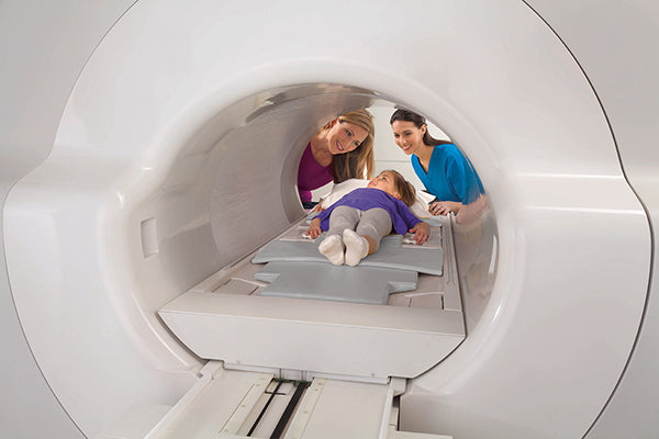
The Fuji Echelon Oval MRI System is a 1.5T wide bore MRI designed for imaging centers, hospitals, and outpatient facilities that need advanced clinical capabilities with improved patient comfort and accessibility. Its 74 cm oval bore provides a spacious scanning environment, while the wide 63 cm patient table and 550 lb weight limit help accommodate a broader range of patient body types.

Built with Fujifilm’s SynergyDrive workflow platform and Workflow Integrated Technology (WIT), the Echelon Oval is designed to streamline patient setup, positioning, scan planning, acquisition, and post-processing. The system also includes patient-focused features such as in-bore lighting and ventilation, feet-first imaging, SoftSound noise reduction, and a mobile table that lowers to 20 inches for easier patient access.

Key Features of the Fuji Echelon Oval

1.5T Wide Bore MRI Design
The Echelon Oval combines 1.5T imaging performance with a spacious 74 cm oval bore, helping improve comfort for anxious, claustrophobic, broad-shouldered, and bariatric patients.

74 cm Oval Bore
The ultra-wide oval bore is designed around the shape of the body to provide more space where patients need it most.

63 cm Wide Patient Table
The system features a wide 63 cm patient table to support better patient accommodation, comfort, and positioning.

550 lb Patient Weight Limit
The detachable mobile table supports patients up to 550 lb and includes vertical power motion for improved accessibility.

SynergyDrive Workflow Technology
SynergyDrive combines Evolution Software and Workflow Integrated Technology to help optimize registration, positioning, scan planning, acquisition, and post-processing.

WIT Monitor and In-Room Controls
The WIT monitor allows technologists to review and update patient information at the gantry, while in-room start, pause, and restart controls help reduce patient anxiety and improve workflow.

WIT Integrated RF Coil System
Integrated head, body, and spine coils work with lightweight anterior coil attachments to improve patient comfort, workflow, and image quality.

SoftSound Noise Reduction
SoftSound technology provides up to 90% reduction in audible noise, helping create a quieter and more comfortable MRI experience.

Digital Drive DX and Optical Data Transmission
Digital Drive DX performs A/D conversion at the gantry, while optical data transmission helps reduce noise and maximize SNR.

Ideal Clinical Applications
The Fuji Echelon Oval MRI system is well suited for facilities performing a wide range of advanced MRI studies, including:

  • Neuro imaging
  • Vascular imaging
  • Orthopedic and MSK imaging
  • Body imaging
  • Breast imaging
  • Prostate imaging
  • Cardiac imaging
  • Pediatric MRI
  • Bariatric and claustrophobic patient exams


Why Choose the Fuji Echelon Oval?

The Fuji Echelon Oval wide bore MRI system gives imaging providers a strong combination of 1.5T performance, advanced workflow automation, and patient-centered design. Its wide oval bore, high-capacity table, integrated RF coil technology, and broad clinical imaging capabilities make it a practical choice for facilities looking to improve patient comfort while maintaining diagnostic confidence.

Key Benefits

  • 1.5T MRI system with 74 cm oval bore
  • Wide 63 cm table with 550 lb weight capacity
  • Patient-friendly design for anxious, bariatric, pediatric, and geriatric patients
  • SynergyDrive workflow tools help streamline exams
  • WIT technology supports efficient setup and positioning
  • SoftSound helps reduce audible scan noise

Specifications

FUJIFILM ECHELON OVAL
1.5T Wide Bore MRI System

Technical Specifications

SYSTEM OVERVIEW

• 1.5T superconducting MRI system
• 74 cm oval wide bore design
• Designed for broad patient access and comfort
• Workflow Integrated Technology / WIT platform
• Advanced image acquisition and processing capabilities
• Built for neuro, orthopedic, body, breast, vascular, and cardiac imaging

MAGNET SYSTEM

• Magnet Type: Superconducting magnet
• Field Strength: 1.5T
• Field Orientation: Horizontal field
• Bore Design: Oval wide bore
• Patient Aperture: 74 cm x 65 cm
• Homogeneity: <0.5 ppm @ 40 cm DSV
• Active magnetic shielding
• Computer-mapped passive shim
• Linear and higher-order per-patient active shim
• HOAST™ Higher Order Active Shim Technology
• Helium Refill Frequency: Once every 6 years with Fujifilm Customer Support Program

5-GAUSS FRINGE FIELD

• Axial: 4.0 m / 13.1 ft
• Radial: 2.5 m / 8.2 ft

PATIENT TABLE & POSITIONING

• WIT Mobile Table
• Patient Weight Capacity: 550 lbs / 250 kg
• Table Width: 63 cm
• Longitudinal Travel: >9 ft / 279 cm
• Vertical Range: 19.7"–33.2" / 50–84 cm
• Feet-first scanning support for any exam
• Class II laser positioning
• Laser Accuracy: ±0.5 mm
• Automatic movement to isocenter

PATIENT AMENITIES

• Two-way intercom
• Technologist alert system
• Adjustable bore illumination
• Adjustable bore ventilation
• Patient pads and immobilization straps
• Arm boards for IV access and patient safety
• WIT gantry monitor for patient, coil, and gating information

GRADIENT SYSTEM

• Peak Gradient Amplitude: 34 mT/m
• Peak Slew Rate: 150 T/m/s
• Active shielding
• Water cooling
• Mechanical gradient sound dampening

RF SYSTEM

• WIT RF System
• Integrated RF receiver coils
• Digital Drive Dx receiver
• Receiver Channels: 16 or 32 channels
• Coil Connection Points: 7
• Ultra-low-noise coil-mounted preamplifiers
• A/D conversion on gantry
• Optical digital transmission to equipment room
• RAPID™ parallel imaging support

RF TRANSMIT

• Two 20 kW RF power amplifiers

AVAILABLE COILS

• WIT Posterior Head / Neck
• WIT Posterior Spine A and B
• WIT Anterior Head
• WIT Anterior Neck
• WIT Anterior Neurovascular
• WIT Torso
• WIT Cardiac
• Shoulder
• Extremity
• Hand / Wrist
• Foot / Ankle
• Flexible Extremity
• Bilateral Breast
• Peripheral Vascular
• Extremity Micro Coil
• QD T/R Body

COMPUTER SYSTEM

• Vertex™ II Computer System
• Core i5 Processor
• RAM: 8 GB
• Monitor: 24" LCD color monitor
• Display Matrix: 1920 x 1200
• Storage Capacity: 320 GB
• Image Storage: Up to 400,000 images at 256 x 256
• DVD Archive Capacity: 4.7 GB
• DVD Storage: Up to 30,000 images at 256 x 256
• CD / DVD writer with auto-launching PC viewer software

SCAN / RECONSTRUCTION ENGINE

• Multiple processors
• Pulse sequence control
• Digital receive
• Image reconstruction
• Post-image reconstruction
• Simultaneous scan and reconstruction

OPERATING SOFTWARE

• Origin™ MRI Operating Software
• Password login security
• Normal and audit user privileges
• Timeout security
• Audit log
• Patient information management
• HIS / RIS registration support
• Automated study ID assignment
• Rapid registration mode
• Patient data correction feature
• Modality Worklist Management
• Fujifilm-provided protocol library
• User-defined protocols
• Graphical scan selection
• Interactive scan control
• Preview window for scan review

IMAGE PROCESSING

• MIP / minIP
• Multi-Planar Reconstruction / MPR
• Vascular Volume Rendering
• Signal Intensity Ratio Map / SIR Map
• Addition / subtraction
• T1 and T2 calculated images
• T2 RelaxMap
• T2* RelaxMap
• Dynamic analysis
• Perfusion analysis
• Diffusion analysis
• ADC trace
• DWI trace
• Tensor / Kurtosis analysis
• Mean Diffusivity / MD
• Fractional Anisotropy / FA
• Spectroscopy analysis
• Filtering

DICOM & NETWORKING

• DICOM 3.0 compliant
• Print
• Query / Retrieve
• Storage
• Storage Commitment
• Modality Worklist Management
• Modality Performed Procedure Step
• IHE Profiles: SWF / PIR, CPI, KIN, Basic Security
• Drag-and-drop archiving / restoring
• Sentinel™ remote customer support
• Remote system and cryogen monitoring
• Remote desktop
• Remote diagnostics
• Remote image review

PULSE SEQUENCES & ACQUISITION

• Spin Echo / SE
• Inversion Recovery / IR
• FLAIR
• STIR
• 2D / 3D Fast Spin Echo / FSE
• Single Shot FSE
• opFSE
• primeFSE
• isoFSE
• Fast Inversion Recovery / FIR
• Double and Triple IR Black Blood
• 2D / 3D Gradient Echo
• Multi-Echo Gradient Echo
• Micro TE
• ADAGE
• 3D GEIR
• FatSep Dixon
• RADAR Motion Compensation
• RAPID Parallel Imaging
• TIGRE™
• SARGE / SG
• Balanced SG / BASG
• Diffusion Weighted Imaging / DWI
• Diffusion Tensor / Kurtosis up to 30 axes
• Perfusion imaging
• BSI
• 2D / 3D TOF
• BeamSat TOF
• VASC-ASL
• FLUTE™ triggered MRA
• TRAQ™ time-resolved MRA
• Phase Contrast MRA
• Non-Contrast MRA

IMAGING PARAMETERS

• 2D Slice Thickness: 0.5–100 mm
• 3D Slice Thickness: 0.05–10 mm
• Field of View: 3–50 cm
• TR Range: 0.9–20,000 ms
• TE Range: 0.25–7,680 ms
• TI Range: 20–8,000 ms
• FSE Inter-Echo Time: 4.4–30 ms
• EPI Inter-Echo Time: 0.4–7 ms
• Signals Averaged: 1–99
• 3D Multi-Slab: 32
• Maximum 2D Slices: 256 at 512 x 512
• Maximum 3D Slices: 512 at 512 x 512
• Acquisition Matrix: Up to 1024 x 1024
• Reconstruction Matrix: Up to 2048 x 2048

GANTRY DIMENSIONS

• Gantry Length: 70.5" / 179 cm
• Gantry Width: 86.6" / 220 cm
• Gantry Height: 86.6" / 220 cm
• Gantry Weight: 9,680 lbs / 4,400 kg

BORE DIMENSIONS

• Oval Bore: 74 cm x 65 cm
• Bore Length: <63" / 160 cm

PATIENT TABLE DIMENSIONS

• Table Length: 89" / 226 cm
• Table Width: 29.5" / 75 cm
• Tabletop Width: 25" / 63 cm
• Minimum Height: 20" / 50 cm
• Maximum Height: 33.2" / 84 cm

SITING REQUIREMENTS

• Typical Scan Room Size: 23.8' x 15.0' / 7.2 m x 4.6 m
• Scan Room Minimum Ceiling Height: 8.7' / 2.65 m
• Equipment Room Size: 13.2' x 9.0' / 4.0 m x 2.7 m
• Control Room Size: 10.1' x 9.0' / 3.1 m x 2.7 m
• RF-shielded scan room required
• RF Noise: <0 dB µV/m from 10–80 MHz
• Power: 3-phase AC 460V / 480V
• Frequency: 50/60 Hz ±1% or less
• Power Capacity: 100 kVA

KEY FEATURES SUMMARY

• 1.5T superconducting MRI
• 74 cm x 65 cm oval wide bore
• 550 lb patient table capacity
• 34 mT/m gradients
• 150 T/m/s slew rate
• 16 or 32 channel receiver
• WIT RF coil system
• RAPID parallel imaging
• RADAR motion compensation
• Vertex II computer system
• Origin MRI operating software
• Advanced neuro, orthopedic, body, breast, vascular, and cardiac imaging support
• DICOM 3.0 connectivity
• Sentinel remote support

FAQs

s the Echelon Oval good for larger or claustrophobic patients?

Yes. The system has a 63 cm patient table, 550 lb weight limit, 74 cm oval bore, in-bore lighting and ventilation, feet-first imaging capability, and SoftSound noise reduction to help improve comfort for larger, anxious, or claustrophobic patients.

Does the Echelon Oval support advanced vascular imaging?

Yes. The brochure highlights vascular tools and examples including FLUTE, VASC-FSE non-contrast MRA, 3D Time-of-Flight, VASC-ASL volume-rendered non-contrast MRA, and Blood Sensitive Imaging for microbleed detection.

What is SynergyDrive on the Echelon Oval?

SynergyDrive is Fujifilm’s workflow platform that combines Evolution Software and Workflow Integrated Technology to streamline patient registration, positioning, scan planning, acquisition, and post-processing.

Additional Information

Condition
New
Brand
Fuji
MPN
Echelon Oval MRI
Specialty
  • Orthopedics
  • Spine
  • Neuro
Country Of Origin
Japan
Tesla Count
1.5 T